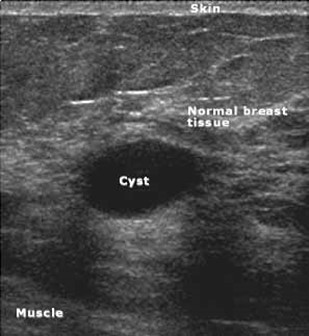
Breast Ultrasound
Breast ultrasound imaging is typically used in conjunction with mammography to further evaluate an area seen on a mammogram. A breast ultrasound may also be
ordered when a patient or physician notices a breast lump or breast pain. Ultrasound is not reliable for screening the breast for abnormalities and does NOT
replace your mammogram. Mammograms are considered the “gold standard” for early detection of breast cancer. Many cancers are not visible on ultrasound. Many
calcifications seen on mammography cannot be seen on ultrasound. Some early breast cancers only show up as calcifications on mammography. An ultrasound is
recommended when a density is seen on a mammogram that requires further evaluation. In some cases, a mammogram will reveal a questionable area in the breast
and ultrasound is used to further evaluate the specific area of interest. Biopsy may be recommended to determine if a suspicious abnormality is cancer or not.

What Should I Expect?
You will change into a gown and lie on an exam table. A gel is applied to your skin to ensure that the transducer (the probe that emits the high-frequency sound waves)
has good contact for sound transmission.
The transducer is placed on your skin and is moved over the area of interest as the technologist records various images. You should experience no pain or
discomfort during the exam.
The radiologist will review your exam and a report of your exam will be sent to your doctor, who will discuss the results with you.